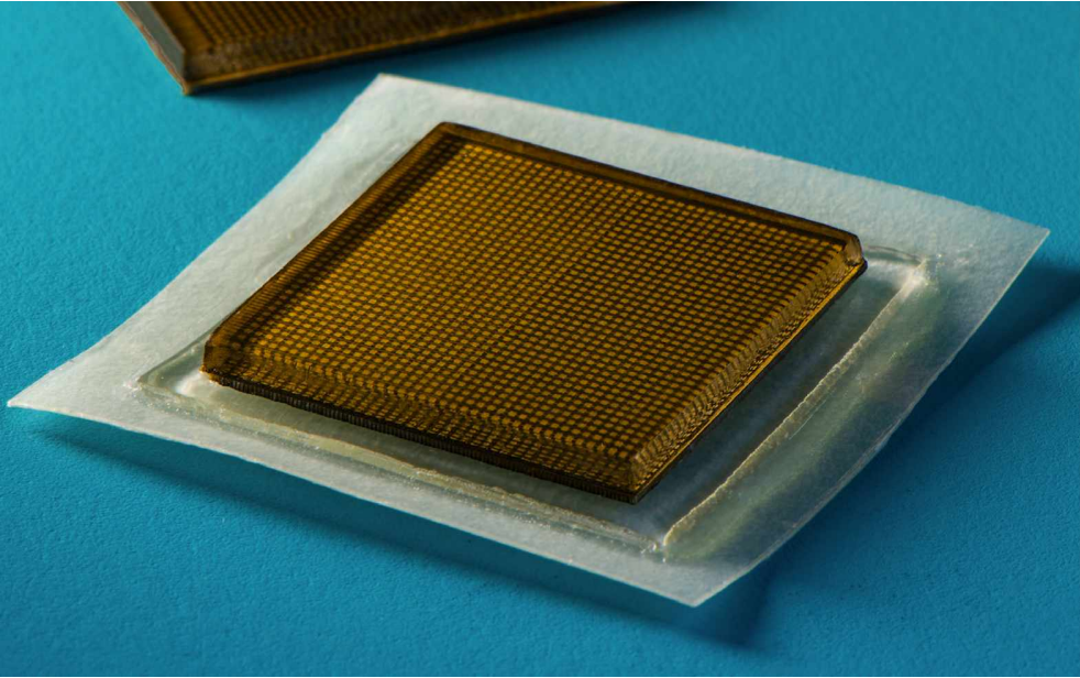
The Massachusetts Institute of Technology (MIT) has combined engineering and medical expertise to create a technology. A team of engineers, at MIT has developed a method that could potentially revolutionize healthcare. This groundbreaking technique involves using a patch that adheres to the skin to enable ultrasound imaging of organs inside the body. In this article we will explore the development, implications and promising future of these ultrasound patches.
The Evolution of Ultrasound Imaging
Ultrasound imaging also known as sonography has come a way since its origins during World War I when it was used for detecting submarines. In the field of medicine, it has been a tool since the 1940s. Most people are familiar with ultrasound as the technology used to capture images of developing babies in the womb. The traditional process involves applying gel on the skin to allow ultrasound waves to penetrate and bounce off structures creating representations of our body’s interior.
Challenges with Patch Designs
For quite some time scientists have aimed to replace ultrasound wands with flexible adhesive patches in order to improve patient comfort and convenience. Earlier designs incorporated sensors, into plastic patches enabling them to move with patients’ movements.
However, these designs encountered problems where the sensors would shift in relation, to each other resulting in images to trying to take a clear photo while running.
MITs Revolutionary Ultrasound Patch
MITs latest advancement is remarkable due to its design. It utilizes an array of sensors that maintain their positions ensuring sharp and clear images. These sensors are attached to a three-layered patch made up of two layers of elastomer surrounding a stretchy hydrogel layer. This water-based material facilitates the transmission of soundwaves allowing the ultrasound waves to effectively penetrate the skin.
The patch has dimensions measuring two centimeters by two centimeters and three millimeters in thickness making it about the size of a postage stamp. To assess its practicality volunteers wore these patches while engaging in activities like sitting, standing, jogging and cycling. Surprisingly the ultrasound scanners remained securely attached throughout these activities. Produced images of internal organs and blood vessels. Notably this patch can provide continuous ultrasound imaging for, up to 48 hours.
Real time Monitoring and Insights
This technological breakthrough provides real time insights into the workings of the body.
Using the captured images researchers observed the dilation and contraction of blood vessels witnessed the heart changing shape during exercise and even detected microdamage, in a volunteer’s muscles while lifting weights. The possibilities for monitoring and understanding the dynamics of the body are practically limitless.
A Wireless Future
Currently the patch is connected to a machine that generates ultrasound images. Nevertheless, the MIT research team has plans for the future. Their goal is to develop a version of the patch that can communicate with devices. By utilizing intelligence (AI) algorithms to analyze data, real time diagnostics and insights can be provided, reducing the necessity for hospital visits.
The Impact on Healthcare
The potential applications of these ultrasound patches are extensive. Hold great promise in revolutionizing healthcare. As these patches continue to advance and become more accessible, they could usher in an era of patient centered care that’s non-invasive. Let’s explore some of the implications:
- Remote Monitoring: Patients with conditions, like heart disease or diabetes could greatly benefit from these patches. They would be able to wear them at home while healthcare professionals continuously monitor their organs and systems.
The patches have the capability to instantly detect any abnormalities and alert medical teams in time ensuring notification of any concerning changes. - Prenatal Care: Expectant parents can greatly benefit from these patches as they offer noninvasive monitoring during pregnancy. This breakthrough technology allows for observation of development, in the womb providing reassurance and enabling early identification of potential issues.
- Early Disease Detection: In terms of disease detection these patches could revolutionize diagnosis by monitoring organs and tissues. This has the potential to be a game changer for diseases like cancer as it allows for real time anomaly detection and facilitates intervention increasing the chances of treatment.
- Postoperative Care: For patients who have undergone procedures involving devices such as stents or plates these patches can provide ongoing monitoring to ensure proper functionality. By detecting any complications or issues with these devices serious complications can be prevented.
- Telemedicine and AI Integration: Furthermore, integrating the patches with devices opens up possibilities for telemedicine applications. Patients can remotely consult with their healthcare providers while AI algorithms analyze data collected by the patches to generate reports. This reduces the need, for frequent doctor visits. Enables patients to receive care comfortably from their homes.
- Reducing Hospital Visits: One of the advantages is that it reduces the need, for time consuming trips to the hospital. Patients can simply apply these patches. Carry on with their lives while their health is being monitored minimizing any disruption or inconvenience.
The Future of Ultrasound Technology
The creation of these ultrasound patches demonstrates the progress made in medical technology. The combination of engineering and healthcare has opened up possibilities that were only found in science fiction. However, this is the beginning. The future holds exciting advancements for ultrasound technology;
- Miniaturization: With technological advancements we can anticipate these patches becoming even smaller and more discreet. This will make them more comfortable for patients to wear while ensuring an data collection process.
- Improved Imaging: Ongoing research aims to enhance the quality and depth of ultrasound scans provided by these patches. This could offer detailed insights into the body’s functions and structures.
- Extended Battery Life: A key focus will be on extending the duration that these patches can function. Lasting batteries or alternative power solutions will be crucial for prolonged monitoring needs associated with chronic conditions.
- Wireless Connectivity: The shift towards patches represents a leap forward, in this field. As the wireless capabilities of patches continue to improve, they will provide convenience by allowing patients to carry on with their daily activities without any limitations.
- AI-Driven Diagnostics: The implementation of intelligence, in diagnostics is set to revolutionize the field. Advanced algorithms will not analyze data. Also offer predictive insights enabling healthcare professionals to anticipate potential issues before they become critical.
In conclusion
The introduction of ultrasound patches at MIT signifies a milestone in healthcare advancement. These patches have the potential to revolutionize how we monitor and manage our wellbeing. From monitoring and early detection of diseases to consultations and reduced hospital visits the possibilities are vast and exciting.
With advancements we can eagerly anticipate further groundbreaking discoveries in ultrasound technology. The vision of a future where healthcare revolves around patients providing convenience and effectiveness is gradually becoming a reality thanks to the minds at MIT and the collaborative efforts between engineering and medical science. It is an exhilarating time for healthcare with a future that looks more promising, than before.
